

Glioblastoma Surgery + NK Cell Immunotherapy
(Investigational approach, not a standard treatment modality)From the First Surgery to Direct Intracavitary Immunotherapy. Glioblastoma is an aggressive brain tumor. It extends microscopically beyond what MRI shows and beyond what surgery can safely remove.
For this reason, when a patient needs surgery, we consider two questions. How much tumor can we safely remove today? And how can the first operation create an opportunity for future treatment directly at the tumor site?
Request a Consultation → Read the 2019 Publication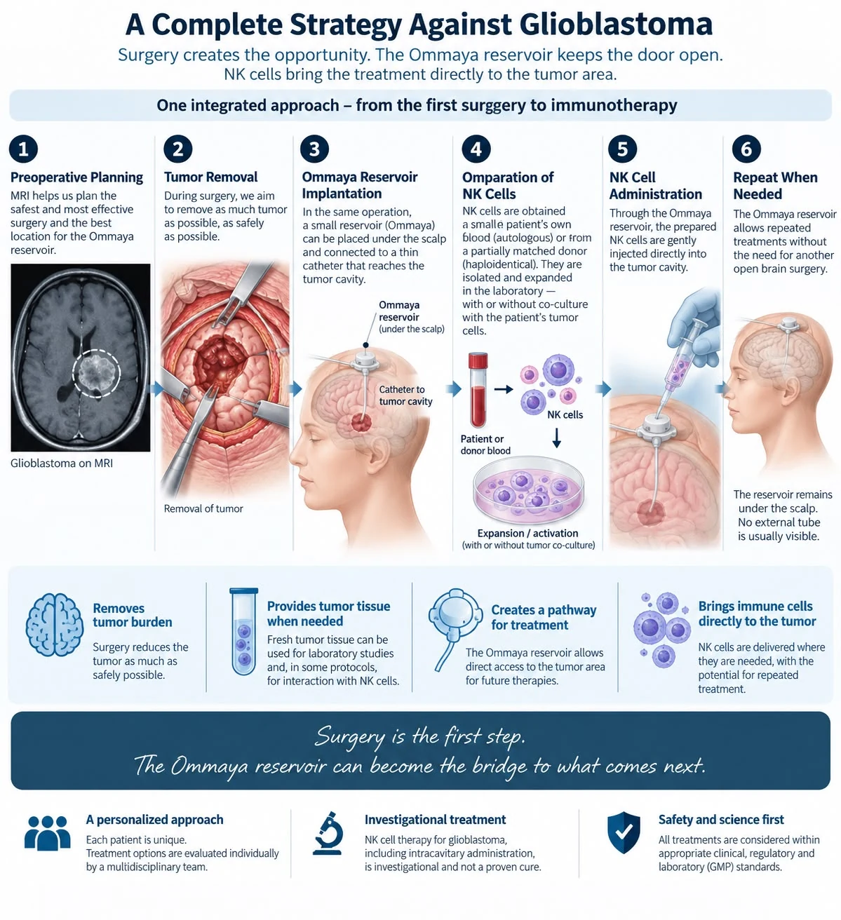
One integrated approach, from the first surgery to immunotherapy. Click to enlarge.
Three Elements, One Strategy
Our approach brings together three elements that connect into a single pathway, planned from the first operation.
Maximal Safe Surgery
We remove as much tumor as we safely can while protecting neurological function.
Ommaya Reservoir
A small chamber implanted under the scalp during the same operation, keeping a pathway open to the tumor area.
NK Cell Immunotherapy
The body's own natural killer cells, prepared in the laboratory and brought directly to where residual tumor cells may remain.
This page describes how these three elements connect into one integrated pathway. It builds on our published 2019 compassionate-use experience with tumor-immunized autologous NK cells. For the general background, see our NK Cell Therapy for Glioblastoma guide.
Maximal Safe Tumor Removal
The first and most important step is neurosurgery. Whenever possible, we aim to remove as much of the visible tumor as we safely can while protecting neurological function.
However, surgery has another important role. It provides fresh tumor tissue. This tissue is essential for pathological and molecular analysis. In addition, it may prove valuable in certain investigational NK-cell strategies.
In our previously published compassionate-use work, we cultured fresh tumor cells. We then used them together with tumor lysate for co-culture with autologous NK cells from the patient's blood.
The first operation therefore matters for more than removing tumor. It can also help prepare the way for what comes next. In this pathway, surgery has three roles:
- Reduce the visible tumor burden as safely as possible
- Provide fresh tumor tissue for diagnosis and molecular studies
- Create the moment to implant the Ommaya reservoir in the same operation
For details on how we approach the operation itself, see glioblastoma surgery in Istanbul.
Implant the Ommaya Reservoir During Surgery
When appropriate, we can implant an Ommaya reservoir during the same operation.
An Ommaya reservoir is a small chamber that sits under the scalp. A thin catheter connects it to the intended intracranial treatment compartment, such as the surgical tumor cavity.
After healing, the reservoir remains beneath the scalp. Consequently, there is normally no external tube.
Why place an Ommaya reservoir during the first surgery?
Because glioblastoma treatment does not necessarily end after surgery removes the tumor. Without such access, delivering treatment repeatedly into the surgical cavity may require another invasive procedure.
The Ommaya reservoir therefore creates a potential long-term access pathway to the tumor area. As a result, we can administer selected investigational treatments intracranially without performing another open brain operation for every treatment.
In simple terms:
- Surgery removes as much tumor as safely possible.
- The Ommaya reservoir keeps a pathway open to the area where residual tumor cells may remain.
For a full explanation of the device, its placement and its risks, see our Ommaya reservoir guide.
Prepare the NK Cells, the Body's Natural Fighter Cells
NK stands for Natural Killer. NK cells are a distinct type of immune cell that circulates naturally in our blood. They belong to the body's own fighter and killer cells, and they can recognize and destroy abnormal cells.
However, a brain tumor presents a fundamental challenge. NK cells in the bloodstream do not normally encounter glioblastoma cells inside the brain the way they encounter abnormal cells elsewhere in the body. In practical terms, the tumor and these circulating immune cells remain largely separated.
Our investigational strategy therefore aims to change this relationship. We want to bring NK cells and glioblastoma cells together so they can interact and fight at the cellular level.
Two Principal Sources of NK Cells
| Source | Where the cells come from | Key consideration |
|---|---|---|
| Autologous NK cells | The patient's own peripheral blood | The laboratory isolates, activates and expands them into a larger population of the patient's own natural killer cells |
| Haploidentical NK cells | A partially HLA-matched donor, often a close family member | Use depends on the individual patient's circumstances and an appropriate clinical and regulatory framework |
NK Cells With or Without Tumor Co-Culture
This distinction is important. Not every NK-cell strategy requires tumor co-culture.
For example, in one approach the laboratory simply isolates, activates and expands the NK cells and prepares them for treatment.
In another investigational approach, the laboratory can additionally co-culture the NK cells with, or otherwise expose them to, the patient's own tumor cells or tumor material. This is possible when viable tumor tissue is available and the laboratory protocol permits. As a result, the patient's immune fighter cells and tumor cells interact directly at the cellular level in the laboratory.
Our original published compassionate-use work followed this latter strategy. We isolated autologous NK cells from peripheral blood and co-cultured them with cultured tumor cells and tumor lysate. We then separated and cultured the tumor-immunized NK cells (Albayrak, J Clin Exp Invest 2019).
In simple terms:
- NK cells are natural fighter cells already circulating in our blood.
- The challenge is bringing these immune cells and the brain tumor together.
- Our methodology aims to let NK cells encounter and fight glioblastoma cells directly at the cellular level, through tumor interaction or co-culture when appropriate, and by bringing prepared NK cells directly to the tumor area.
Bring the NK Cells Directly to the Tumor Area
Once the laboratory has prepared the NK cells, the Ommaya reservoir then provides a potential route for direct intracavitary administration.
NK cells given through the bloodstream must find their own way to the brain tumor. Instead of relying only on that route, our concept is simple: bring the immune cells directly to where the tumor cells remain.
Through the previously implanted catheter, the Ommaya reservoir can potentially deliver prepared NK cells into the intended intracranial compartment. This is the central rationale for combining neurosurgery and cellular immunotherapy from the beginning.
Similarly, the same reservoir-based route also serves other intracavitary treatments. We explain the general principle in intracavitary chemotherapy for brain tumors. The key points of this step:
- NK cells reach the tumor area itself, where they are needed
- The administration does not require a new open brain operation
- The approach targets the intracranial compartment chosen at the time of surgery
Potential for Repeated Treatment
The Ommaya reservoir remains beneath the scalp after surgery. Therefore, when clinically and regulatorily appropriate, it may provide access for repeated intracranial treatments. Each treatment then does not require a new open brain operation.
This creates a potential treatment pathway:
- Brain tumor surgery: maximal safe tumor removal, with fresh tumor tissue obtained when needed
- Ommaya reservoir implanted during the same surgery
- NK cell source: patient blood (autologous NK cells) or donor blood (haploidentical NK cells)
- NK cell isolation, activation and expansion, with or without tumor co-culture
- NK cells delivered through the Ommaya reservoir: direct intracavitary access to the tumor area
- Potential for repeated treatment through the same reservoir
Why We Plan This From the First Surgery
For an appropriately selected patient, the first operation can therefore serve three complementary purposes:
- Reduce tumor burden. Remove as much tumor as safely possible.
- Obtain fresh tumor tissue. Provide tissue for diagnosis and molecular studies and, when appropriate, for potential tumor and NK cell interaction or co-culture.
- Create a pathway for future treatment. Implant an Ommaya reservoir during the same operation to provide potential direct access to the intracranial tumor area.
For this reason, we ideally consider these possibilities before the first operation, rather than only after surgery has taken place. Patients who are evaluating their options may also find our overview of treatment approaches and immunotherapy for brain tumors helpful for context.
From Surgery to Cellular-Level Treatment
The philosophy is straightforward:
- Remove what can safely be removed.
- Preserve fresh tumor tissue when needed.
- Create access to the tumor area.
- Prepare the body's natural fighter cells.
- Bring those fighter cells and tumor cells together.
Surgery is the first step. The Ommaya reservoir can become the bridge to what comes next.
Our Previous NK-Cell Experience: The 2019 Case
Our published 2019 compassionate-use experience investigated tumor-immunized autologous NK-cell therapy in a child with a malignant pontine glioma. We co-cultured tumor cells and tumor lysate with autologous NK cells isolated from peripheral blood. In that published case, we administered the prepared NK cells intravenously, under authorization from the Republic of Turkey Ministry of Health.
Publication
Albayrak SB. Tumor Immunized Autologous Natural Killer Cell (NK) Therapy/Compassionate Use. Journal of Clinical and Experimental Investigations. 2019;10(3):em00727. Letter to the Editor, open access. DOI: 10.5799/jcei/5858
- Received / accepted: 15.06.2019 / 29.06.2019
- Setting: Department of Neurosurgery, Istanbul Aydin University, with ethics committee approval and Ministry of Health authorization
- Route in the published case: intravenous administration of the prepared NK cells
- Declarations: the authors report no conflicts of interest; no financial support was received
- Reference cited: Pollack IF, Agnihotri S, Broniscer A. Childhood brain tumors: current management, biological insights, and future directions. J Neurosurg Pediatr. 2019;23:261-273.
What the Published Case Involved
A four-year-old boy was referred with a radiological diagnosis of diffuse infiltrative pontine glioma (DIPG). As the publication notes, this tumor carries a dismal prognosis, with a mean survival of around one year despite all available treatment modalities (Pollack et al., 2019). Before referral, he had received radiotherapy and a ventriculoperitoneal shunt at a health facility in Europe.
On admission, seven months after diagnosis, the child was tracheostomized and conscious. He was quadriparetic, with bilateral abducens and lower cranial nerve paresis. We decided to operate in order to debulk the necrotic pontine mass and to verify the radiological diagnosis histopathologically.
In addition, after ethics committee approval from Istanbul Aydin University, we planned to co-culture the tumor cells with NK cells isolated from the patient's peripheral venous blood. The aim was to investigate the immunization and potentiation of autologous NK cells against glioma cells in vitro.
January 2019
Partial removal of the pontine mass via a telo-velar approach under neurophysiological monitoring. No additional postoperative deficits; the unilateral abducens nerve recovered. Tumor specimens went to the tissue culture laboratory. Routine histopathology officially reported the lesion as DIPG (Grade 4 astrocytoma).
March 2019, two months after surgery
After serial tumor cell culture passages, tumor cells and tumor lysate were co-cultured with the isolated autologous NK cells. The tumor-immunized NK cells were then separated and cultured separately.
April 2019
The patient deteriorated because of increased pons edema, decompensated hydrocephalus and a concomitant pulmonary influenza B infection. We placed another ventriculoperitoneal shunt and admitted him to the pediatric intensive care unit. Following antiviral therapy, resolution of hydrocephalus and respiratory support, his level of consciousness improved with limited cooperation.
15 and 23 May 2019
With approval of the clinical trial by the Republic of Turkey Ministry of Health, we administered autologous NK cells intravenously. In each session: 300,000 NK cells in 50 cc physiological saline over 30 minutes.
One week after the second infusion
Post-trial cranial CT showed no increase in pons edema and, on the contrary, slightly more discrete borders in the tumor necrosis.
As of publication, we observed no early or late adverse reactions, including infection, anaphylaxis or thrombosis.
The author's conclusion in the publication: application of autologous NK cell therapy in gliomas and most other solid tumors is still a "no man's land". Therefore, neurosurgeons and clinicians should take the initiative and cooperate closely with basic science researchers to develop a definitive cure in gliomas. In this context, the author found it useful and encouraging to announce this clinical trial.
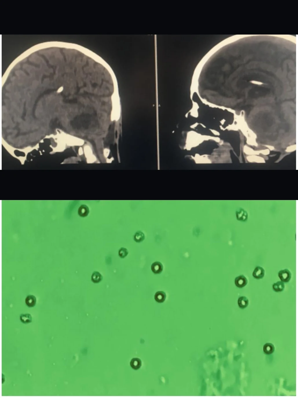
Figure 1 from the publication. Upper panel: cranial CT scans, left image recent and right image one week earlier. Lower panel: light microscopy of immunized NK cells. Reproduced from the open-access article; scanner header text has been removed to protect patient privacy.
How the present concept differs. The 2019 case used intravenous administration, whereas the present concept relies on direct intracavitary administration through an Ommaya reservoir. This is a distinct investigational strategy, so readers should not confuse the two. You can find our other published work under scientific publications and case studies.
Frequently Asked Questions
What is an Ommaya reservoir?
An Ommaya reservoir is a small chamber that sits under the scalp. A thin catheter connects it to the intended intracranial treatment compartment, such as the surgical tumor cavity. After healing, the reservoir remains beneath the scalp, and there is normally no external tube.
Why is the Ommaya reservoir implanted during the first surgery?
Because glioblastoma treatment does not necessarily end after tumor removal. Implanting the reservoir in the same operation creates a potential long-term access pathway to the tumor area. As a result, we can administer selected investigational treatments intracranially without another open brain operation for every treatment.
What are NK cells?
NK stands for Natural Killer. NK cells are a distinct type of immune cell that circulates naturally in the blood, and they can recognize and destroy abnormal cells. However, circulating NK cells do not normally encounter tumor cells inside the brain the way they encounter abnormal cells elsewhere.
Is tumor co-culture required for every NK-cell strategy?
No. In one approach, the laboratory simply isolates, activates and expands the NK cells. In another investigational approach, the laboratory can additionally co-culture the NK cells with, or otherwise expose them to, the patient's own tumor cells or tumor material, when viable tissue is available and the protocol permits.
Where do the NK cells come from?
There are two principal sources. Autologous NK cells come from the patient's own peripheral blood, whereas haploidentical NK cells come from a partially HLA-matched donor, often a close family member. Their use depends on the individual patient's circumstances and an appropriate clinical and regulatory framework.
What did the 2019 published case show?
In a four-year-old child with DIPG, we co-cultured tumor cells and tumor lysate with autologous NK cells, then administered 300,000 tumor-immunized NK cells intravenously on two occasions under Ministry of Health authorization. As of publication, there were no early or late adverse reactions, and cranial CT one week later showed no increase in pons edema and slightly more discrete tumor necrosis borders.
How does the present concept differ from the 2019 case?
In the 2019 case, we administered the prepared NK cells intravenously. The present concept of direct intracavitary administration through an Ommaya reservoir is a distinct investigational strategy. Therefore, readers should not confuse it with that intravenous approach.
Is NK-cell therapy a proven cure for glioblastoma?
No. NK-cell therapy for glioblastoma remains investigational, including autologous and haploidentical NK cells, tumor co-culture strategies and direct intracavitary administration through an Ommaya reservoir. It is not established standard-of-care treatment, and it has not been proven to cure glioblastoma. Every patient requires individual evaluation within appropriate clinical, ethical, regulatory, laboratory (GMP) and safety frameworks.
Important Information
NK-cell therapy for glioblastoma remains investigational. This includes autologous NK cells, haploidentical NK cells, tumor co-culture strategies and direct intracavitary administration through an Ommaya reservoir. It is not established standard-of-care treatment, and it has not been proven to cure glioblastoma.
Every patient requires individual evaluation. In addition, any cellular treatment must take place within appropriate clinical, ethical, regulatory, laboratory/GMP and safety frameworks.
Medical Disclaimer: This page provides general educational information only. It does not constitute medical advice and cannot substitute for professional diagnosis or treatment.
Please consult Prof. Dr. Serdar Baki Albayrak or another qualified physician directly regarding any diagnosis or treatment decision. For a personal evaluation, contact our clinic.
Discuss Whether This Pathway Fits Your Situation
Every glioblastoma patient is different. Our team evaluates surgery, Ommaya reservoir placement and NK cell immunotherapy individually, within the appropriate clinical and regulatory framework. Therefore, contact us to learn whether this integrated approach may be appropriate for you or your loved one.
Schedule a Consultation →