

Pilomyxoid Astrocytoma: Prognosis, Survival Rate and Treatment
Pilomyxoid astrocytoma is a rare variant of pilocytic astrocytoma. It mainly affects infants and young children. Its prognosis is less favorable than classic pilocytic astrocytoma. Specifically, it recurs more often and progresses sooner. Nevertheless, most children survive many years, and newer molecular treatments are improving the outlook. This guide explains what the diagnosis means and what the survival data show. It also covers how doctors treat it.
Request a Consultation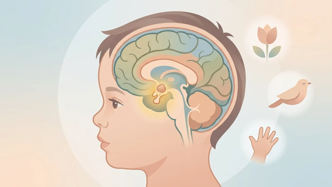
At a Glance
Terminology note: The WHO 2021 classification no longer lists pilomyxoid astrocytoma as a separate grade. Instead, it treats the pilomyxoid pattern as a variant of pilocytic astrocytoma. Older reports describe it as a WHO grade 2 tumor. So you may see both terms in medical records.
What Is a Pilomyxoid Astrocytoma?
A pilomyxoid astrocytoma is a low-grade glioma. Pathologists first described it as a distinct entity in 1999. It shares its origin and, moreover, its BRAF biology with pilocytic astrocytoma. However, its microscopic appearance and its clinical behavior differ. Therefore, doctors treat it as a separate group for prognosis.
Under the microscope, the tumor shows uniform bipolar cells in a myxoid, mucus-like background. The cells often arrange themselves around blood vessels, in an angiocentric pattern. In contrast to classic pilocytic astrocytoma, it lacks Rosenthal fibers and eosinophilic granular bodies.
Who Develops It and Where It Grows
Pilomyxoid astrocytoma mainly affects infants and toddlers, especially those under two. The median age at diagnosis is about 18 months. Most patients, in fact, are under four years old. Occasionally, older children and young adults receive the diagnosis.
The tumor generally grows in the hypothalamus and optic chiasm. These structures sit deep at the base of the brain. This location explains much of its prognosis. Surgeons rarely can remove it completely without harming vision or hormone function. Less often, the tumor appears in the thalamus, cerebellum, brainstem, or spinal cord.
How It Differs From Pilocytic Astrocytoma
The differences between the two tumors affect both treatment and prognosis, so they matter. The table below summarizes them.
| Feature | Pilomyxoid Astrocytoma | Classic Pilocytic Astrocytoma |
|---|---|---|
| Typical age | Infants, median about 18 months | Children 5 to 15, some adults |
| Typical location | Hypothalamus and optic chiasm | Cerebellum, hemispheres |
| Rosenthal fibers | Absent | Present |
| Myxoid background | Prominent | Minimal |
| Complete resection | Rarely possible | Usually possible |
| Recurrence | Common | Uncommon after complete removal |
| Spread through CSF | Reported in a minority | Very rare |
For a full picture of the classic form, read our guide to grade 1 pilocytic astrocytoma.
Symptoms in Infants and Young Children
Symptoms generally reflect the hypothalamic and chiasmatic location. Infants cannot describe what they feel. Instead, parents and pediatricians notice changes in growth, feeding, and vision. Common presentations include:
- Failure to thrive: Poor weight gain despite normal or increased appetite, sometimes called diencephalic syndrome
- Vision problems: Nystagmus, poor visual tracking, or a squint
- Hydrocephalus: Rapid head growth, vomiting, irritability, and drowsiness
- Hormone disturbance: Early puberty, abnormal thirst, or altered temperature regulation
- Developmental delay: Slower milestones than expected
Consequently, any infant with unexplained failure to thrive and eye movement abnormalities needs a brain MRI without delay.
Diagnosis and MRI Features
MRI is the first and, indeed, the most important test. A pilomyxoid astrocytoma generally appears as a large, mostly solid mass in the hypothalamic region. It enhances brightly and evenly with contrast. Unlike classic pilocytic astrocytoma, it rarely shows the cyst-with-nodule pattern. In addition, bleeding inside the tumor occurs more often than in the classic form.
The tumor sits deep in the brain. Therefore, surgeons obtain tissue through a planned resection or a targeted biopsy. Pathologists then confirm the diagnosis and, additionally, test for BRAF alterations. Consequently, the diagnostic work-up includes MRI of the spine as well. This checks for spread through the cerebrospinal fluid.
Pilomyxoid Astrocytoma Prognosis: The Numbers
The prognosis for pilomyxoid astrocytoma is more guarded than for classic pilocytic astrocytoma. However, it is far from hopeless. The most cited comparison comes from a 2004 series of hypothalamic and chiasmatic tumors. In that study, mean progression-free survival reached about 25 months for pilomyxoid tumors. For pilocytic tumors, it reached about 163 months. Mean overall survival was roughly 63 months versus 213 months.
Those figures date from an era before targeted therapy and before modern chemotherapy protocols. More recent series report better outcomes. Many children remain alive and stable a decade after diagnosis. Therefore, families should read the older numbers as a description of relative risk. They are not a fixed expectation.
Survival Rates Compared
Direct survival percentages for pilomyxoid astrocytoma vary widely between studies. The tumor is rare, so each series is small. The table below gives the general pattern.
| Measure | Pilomyxoid Astrocytoma | Pilocytic Astrocytoma |
|---|---|---|
| Recurrence or progression | Majority of patients over time | About 10 percent after complete removal |
| Mean progression-free survival | About 2 years (older series) | Over 10 years |
| 5-year overall survival | Roughly 60 to 85 percent across series | 95 to 97 percent |
| Long-term survival | Common with ongoing treatment | Usual, often without treatment |
Our article on pilocytic astrocytoma survival rate and life expectancy gives the full data for the classic form.
What Shapes the Prognosis
Several factors influence how a pilomyxoid astrocytoma behaves in an individual child. They include:
- Extent of resection: Greater removal links with longer progression-free survival. Complete removal, however, is rarely safe in the hypothalamus.
- Age at diagnosis: Infants under one year have the most difficult course. Their bodies tolerate chemotherapy less well.
- Location: Tumors outside the hypothalamus, for example in the cerebellum, allow more complete surgery. Consequently, they do better.
- Molecular profile: Many tumors carry a KIAA1549-BRAF fusion. This now opens the door to targeted therapy.
- CSF dissemination: Spread through the cerebrospinal fluid at diagnosis predicts a harder course.
Recurrence and CSF Spread
Recurrence is, above all, the main challenge in pilomyxoid astrocytoma. Surgeons usually leave tumor behind, so regrowth is common. It tends to occur within the first two to three years. In addition, a minority of tumors seed the cerebrospinal fluid. They then appear at distant sites along the brain and spine.
For this reason, doctors image the entire neuroaxis, both brain and spine. They do so at diagnosis and during follow-up. Early detection of spread changes the treatment plan, so this imaging is not optional.
Treatment: Surgery, Chemotherapy, and Targeted Therapy
Treatment follows the same ladder as pilocytic astrocytoma. However, it relies more heavily on medical therapy. Surgery comes first. It relieves pressure, obtains tissue for diagnosis and molecular testing, and removes as much tumor as is safe. Hydrocephalus, in addition, often requires a shunt or endoscopic third ventriculostomy.
Because residual tumor is the rule, most children receive chemotherapy after surgery. Carboplatin and vincristine form the most common first-line regimen. Weekly vinblastine offers a well-established alternative. Doctors avoid radiation in this age group whenever possible. It harms the developing brain and endocrine system.
Targeted therapy represents the most important recent advance. Tovorafenib received FDA accelerated approval in 2024 for relapsed pediatric low-grade glioma with a BRAF fusion or V600 mutation. Similarly, dabrafenib plus trametinib has approval for BRAF V600E tumors. Consequently, molecular testing of every pilomyxoid astrocytoma is now standard. Our summary of pilocytic astrocytoma treatment guidelines covers these drugs in detail.
Does the Tumor Change Over Time?
Interestingly, yes, in some cases. Several reports describe pilomyxoid astrocytomas that change on recurrence. They show the classic features of pilocytic astrocytoma, including Rosenthal fibers. Pathologists call this maturation, specifically. It suggests that the pilomyxoid pattern represents an early, immature stage of the same tumor.
Maturation matters for prognosis because a tumor that matures tends to behave more indolently afterward. Therefore, doctors re-examine pathology at each recurrence and adjust the outlook accordingly.
Follow-Up and Long-Term Care
Children with pilomyxoid astrocytoma generally need lifelong follow-up by a multidisciplinary team. MRI of the brain and spine typically occurs every three months during treatment. Afterward, it occurs every six months. In addition, pediatric endocrinologists monitor growth and hormones, and ophthalmologists track vision.
Developmental support, nutrition, and school planning also form part of care. Most children survive many years. Therefore, quality of life deserves as much attention as tumor control. Families benefit from a single coordinating clinician who keeps the whole picture in view.
Questions to Ask Your Child's Treatment Team
Generally, families cope better when they understand the plan. Therefore, we encourage parents to bring a written list of questions to each consultation. Useful questions include:
- How much of the tumor can you remove safely, and what would complete removal risk?
- Did the tumor undergo BRAF testing, and what did the result show?
- Does my child need spine imaging, and how often will scans repeat?
- Which chemotherapy regimen do you recommend first, and why?
- When would a targeted drug such as tovorafenib become an option?
- Who coordinates hormone, vision, and developmental follow-up?
Clear answers to these questions reveal the team's real experience with this rare tumor. Moreover, they help families compare opinions if they seek a second opinion.
Expert Insight: Prof. Dr. Serdar Baki Albayrak
Rare Tumors Need Experienced Teams
Prof. Dr. Serdar Baki Albayrak has treated hypothalamic and chiasmatic tumors in infants and children for more than 27 years. For pilomyxoid astrocytoma, he emphasizes safe surgery that preserves vision and hormones. Molecular testing then unlocks targeted therapy.
"With a pilomyxoid astrocytoma, heroic surgery can do harm. The right operation relieves pressure, secures the diagnosis, and protects the child's future. Then modern drugs do the rest of the work."
Prof. Dr. Serdar Baki Albayrak
Ask About Your Child's Treatment Options
We review MRI, pathology, and molecular findings. Then we explain the safest surgical and medical plan for a pilomyxoid astrocytoma.
Schedule a ConsultationFrequently Asked Questions
What is the prognosis for pilomyxoid astrocytoma?
The prognosis is more guarded than for classic pilocytic astrocytoma. Recurrence is more frequent, and progression comes sooner. Even so, most children survive many years. Moreover, outcomes have improved with modern chemotherapy and BRAF-targeted therapy.
What are the survival rates for pilomyxoid astrocytoma?
Reported 5-year survival ranges roughly from 60 to 85 percent across small series, overall. An influential 2004 study found mean overall survival of about 63 months. For pilocytic tumors, it found 213 months. Newer series, however, report better results.
Is pilomyxoid astrocytoma the same as pilocytic astrocytoma?
They are close relatives, yet they differ. Pilomyxoid astrocytoma affects younger children and grows mainly in the hypothalamus. It lacks Rosenthal fibers and recurs more often. The WHO 2021 classification treats it as a variant within the pilocytic astrocytoma family.
Can doctors cure pilomyxoid astrocytoma?
Cure is possible when the tumor sits in a location that allows complete removal. The cerebellum is one example. In the hypothalamus, doctors aim for long-term control rather than cure. They use surgery, chemotherapy, and targeted drugs in sequence.
Does pilomyxoid astrocytoma spread?
A minority of tumors spread through the cerebrospinal fluid. They reach other parts of the brain or spine. This is why doctors image the whole neuroaxis at diagnosis and during follow-up. Spread outside the nervous system, in contrast, essentially never occurs.
Which treatment works best for a pilomyxoid astrocytoma?
Doctors generally start with safe surgery and then give chemotherapy, usually carboplatin and vincristine. For tumors with a BRAF fusion or mutation, targeted drugs offer an effective option, especially after relapse. Examples include tovorafenib and dabrafenib plus trametinib.
A Harder Course, But a Long Road
Pilomyxoid astrocytoma carries a more guarded prognosis than its classic cousin. Its location, its age group, and its tendency to recur explain the difference. However, most children live for many years. Moreover, BRAF-targeted therapy has strengthened every step of treatment. The older survival figures describe relative risk, not the ceiling of what is possible today.
Therefore, families should seek a center with experience in infant brain tumors. Access to molecular testing matters too. Learn about our approach to brain tumor surgery in Istanbul. Also, explore our overview of treatment approaches.
Talk to Our Team
We explain the diagnosis, the treatment sequence, and the realistic outlook. We use plain language.
Contact Us TodayDisclaimer: This page is provided for informational purposes only and does not constitute medical advice. Statistics represent published group averages and cannot predict any individual outcome. Always consult Prof. Dr. Serdar Baki Albayrak or another qualified physician directly regarding any medical condition, symptoms, or treatment decisions.
Discuss Your Case With Our Team
Send your MRI report and pathology details for a structured, honest opinion from an experienced brain tumor surgeon in Istanbul.
Request a Consultation